Gum recession: when is a gum graft worthwhile?
Receding gums do not always call for a graft. The decision depends on the cause, the bone between the teeth, the thickness of the gum and what bothers you. This page explains the signs, the examination, the criteria for deciding, the gum graft techniques used at Dr Hazout's practice in Levallois-Perret and what the studies suggest you can expect.
Receding gums and gum recession: what are we talking about?
Patients describe "receding gums" or a tooth that seems to be "losing its gum": the tooth looks longer and its root becomes visible. The medical term is gingival recession: the gum margin has moved below the junction between the enamel and the root, leaving part of the root exposed. The root is covered by a softer tissue than enamel and becomes exposed to cold, wear and decay.
Receding gums actually cover two situations. In the first, the gum moves down on a tooth whose support between the teeth is intact: this is an isolated recession, often linked to brushing or thin gum tissue. In the second, the gum follows bone destroyed by periodontitis: recession then affects several teeth and comes with pockets, bleeding and sometimes loose teeth. The approach is not the same, and this is the first point the examination must settle (see recognising periodontitis).
Which signs point to a recession?
Recession develops slowly and often goes unnoticed at first. The signs that bring people to the practice fall into four groups.
- sensitivity to cold, air or sweet food, located at the neck of one or more teeth: the exposed root has no enamel to protect it;
- a tooth that looks longer than its neighbours, or a gum line that is no longer level with the teeth next to it;
- a visible root, often more yellow than the enamel, sometimes with a notch at the neck or a cavity;
- discomfort when smiling or brushing: the area hurts when the brush passes, or the gum seems to be missing in front of a tooth.
Bleeding, redness or swelling alongside these signs point instead to inflammation, gingivitis or periodontitis, which has to be treated before a graft is considered. Conversely, an isolated recession on healthy gum does not bleed and does not hurt apart from sensitivity to cold.
Why do gums recede?
Several factors are often associated with recession developing or worsening, and they usually combine. The background first: thin, shallow gum tissue (a thin gingival phenotype) and very thin or even absent bone over the root. On such fragile ground, a modest insult is enough to make the gum retreat.
The most frequent insults are forceful or horizontal brushing with a hard brush, orthodontic movement that pushes the tooth out of its bony envelope, a lip or cheek frenum that pulls on the gum with every movement, a lip or tongue piercing, and an overhanging restoration or a crown whose edge irritates the gum. Periodontitis, finally, destroys bone and attachment; the gum follows, and recession then affects the visible surfaces as well as the spaces between the teeth.
Smoking and poor plaque control do not explain a recession on their own, but they complicate the healing of a graft and weigh in the decision.
What the periodontist examines before deciding
The examination first looks for the cause, then assesses what can be achieved. Dr Hazout measures the height of the recession, the thickness of the gum and the height of keratinised gum (the firm, attached gum, as opposed to the mobile lining mucosa beyond it). He looks at the state of the exposed root (wear, notch, decay), the position of the frenum and the brushing technique, and notes whether the area shows when you smile. X-rays and pocket depth measurements define the bone present between the teeth, because that is what determines the outcome [1].
The 2018 international classification adopts the Cairo classification, based on the attachment (support) between the teeth on either side of the recession [1]:
| Type | What it means | Chance of covering the root |
|---|---|---|
| RT1 | No loss of support between the teeth | Complete coverage possible |
| RT2 | Loss of support between the teeth, less than or equal to that on the visible surface | Coverage often partial |
| RT3 | Loss of support between the teeth greater than on the visible surface | Complete coverage generally not possible |
This classification gives a prognosis even before a technique is chosen: in RT1, full root coverage can be hoped for; in RT2, coverage is often partial; in RT3, the height lost between the teeth is not rebuilt, and the aim becomes thickening the gum and stabilising the situation rather than covering the root [1].
Is a graft needed?
A stable recession that is barely visible, not sensitive, on healthy gum that is easy to clean, can simply be monitored. The cause is corrected first: brushing technique, a soft toothbrush, control of periodontitis through periodontal treatment. A baseline measurement, repeated at each check-up, shows whether the recession is progressing.
A graft is more likely to be discussed when:
- the recession gets worse from one measurement to the next;
- sensitivity to cold persists despite desensitising toothpastes;
- the exposed root is wearing away or decaying;
- the gum is too thin or absent and brushing becomes painful;
- orthodontic treatment is planned in a fragile area;
- the appearance bothers you.
The choice also takes into account what does not depend on the technique: a recession caused by unstabilised periodontitis is not grafted, and fragile gum in a smoker heals less well. When a tooth is too damaged or too loose to be kept, the question shifts to replacing it, which is covered on the Implants page.
Gum graft techniques
The term gum graft, or gingival graft, covers several procedures. All of them pursue one of two aims, or both: recreating a band of firm gum where it is missing, or covering an exposed root. The grafted tissue most often comes from the patient's own palate, whose thick mucosa heals over after harvesting.
Coronally advanced flap. The neighbouring gum is lifted, moved up over the root and stitched. It suits shallow recessions where the gum is thick enough.
Connective tissue graft. A thin graft of connective tissue (the deep layer of the mucosa) is taken from the palate through a small incision, then slipped under the gum, which covers it. It thickens the gum and makes root coverage more stable. It is the reference technique in root coverage studies [2].
Tunnel technique. Instead of cutting the papillae (the gum between the teeth), a "tunnel" is created under the gum of several teeth and the graft is slipped into it. It is often used for multiple recessions and limits visible incisions.
Free gingival graft from the palate. A graft including the surface layer of the palate is placed on the area to be reinforced. Its main purpose is to recreate a band of firm gum where it is missing, rather than to cover the root. Its colour may remain different from the surrounding gum, which tends to reserve it for less visible areas.
Strengthen or cover. In a deep recession with no firm gum, the two aims can follow one another: strengthen first, then cover at a later stage. The clinical case below illustrates this situation.
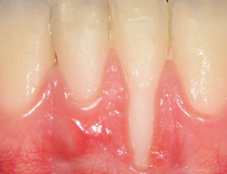
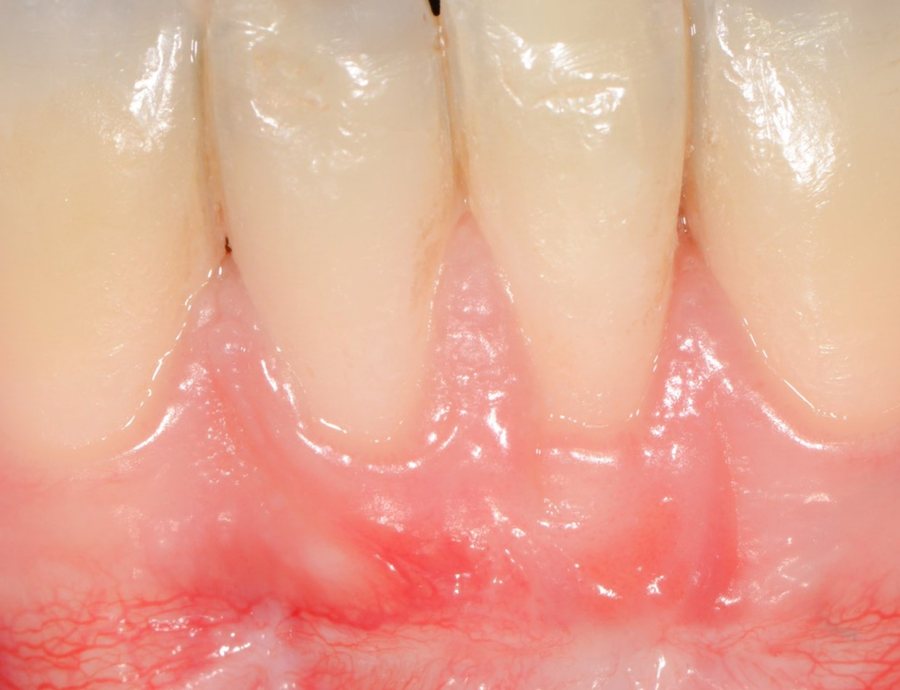
Biomaterials (enamel matrix derivative, soft-tissue substitutes) can sometimes avoid or reduce harvesting from the palate; their value is assessed case by case [2].
Grafts are part of a wider approach to the balance between gums and teeth, set out on the Gum aesthetics page.
The practical arrangements, the course of treatment and aftercare instructions are explained at the practice, according to your situation.
What the studies suggest you can expect
The 2018 Cochrane review analysed 48 randomised trials (1,227 participants) [2]. Several techniques can cover roots: a coronally advanced flap alone or combined with a biomaterial, or a connective tissue graft. A connective tissue graft combined with a coronally advanced flap provides more keratinised gum than the alternatives compared. But the quality of the evidence is low to very low, and the authors remain uncertain which technique is most effective.
For the tunnel technique, a 2018 meta-analysis reports mean root coverage of about 83% for single recessions and 88% for multiple recessions; with the same graft, the coronally advanced flap more often achieved complete coverage than the tunnel [3].
The limits, and why follow-up matters
A graft does not put gum back where the bone between the teeth has gone: in RT2 and especially RT3, coverage remains partial, and nobody can say in advance how much height will be regained. Nor does it correct the cause. If brushing remains traumatic, if a frenum keeps pulling or if orthodontic movement moves the tooth out of its bone, recession can return, on the grafted tooth or on a neighbour.
Follow-up therefore has two purposes: checking that the grafted gum stays stable, and keeping the cause under control. For a patient treated for periodontitis, this check is part of periodontal maintenance visits. For others, a regular measurement of the recession and a look at the brushing technique are usually enough. Recessions that are part of a wider loss of support belong to periodontitis treatment or periodontal regeneration, not to an isolated graft.
Frequently asked questions
Can the gum grow back on its own?
No. Once the cause is corrected, a recession may stabilise, but the gum does not spontaneously move back up over the root.
Is a graft permanent?
Results are generally stable when the cause, such as traumatic brushing, has been corrected. Thicker gum is more resistant, but follow-up is still needed.
Can harvesting from the palate be avoided?
Sometimes, with a flap alone or a substitute. These options suit certain situations; they are discussed according to the type of recession and gum thickness.
Should recession be treated before orthodontics?
Not routinely. If the gum is very thin on a tooth that will be moved outwards, prior reinforcement may be discussed with the orthodontist.
How much does a gum graft cost?
The amount depends on the number of teeth and the technique; a written estimate is given after the examination. The practice's fee ranges are on the Fees page.
References
- Cortellini P, Bissada NF. Mucogingival conditions in the natural dentition: Narrative review, case definitions, and diagnostic considerations. J Clin Periodontol. 2018;45 Suppl 20:S190-S198. DOI
- Chambrone L, Salinas Ortega MA, Sukekava F, Rotundo R, Kalemaj Z, Buti J, Pini Prato GP. Root coverage procedures for treating localised and multiple recession-type defects. Cochrane Database Syst Rev. 2018;10:CD007161. DOI
- Tavelli L, Barootchi S, Nguyen TVN, Tattan M, Ravidà A, Wang HL. Efficacy of tunnel technique in the treatment of localized and multiple gingival recessions: A systematic review and meta-analysis. J Periodontol. 2018;89(9):1075-1090. DOI
Patient pathway