Bone grafting before an implant: when, and which techniques
An implant must be surrounded by bone along its whole length. When bone is lacking, several techniques can rebuild it. They are not equally suited to every situation: everything depends on the shape and size of the defect. Here is how bone loss is assessed, how the technique is chosen, what is used, the principle of the operation, and the risks and limitations you should know about.
Why is bone missing, and how is it measured?
The main cause is resorption after extraction. With no root to support, the ridge becomes thinner: on average 3.8 mm of width and 1.2 mm of height are lost in six months according to a systematic review [1]. Long-standing periodontitis is the other major cause: the infection has destroyed bone around the roots even before the tooth was lost, and the defect is then often deeper and more irregular. An infection at the tip of a root, a cyst, a fracture or trauma can also have removed bone. In the upper jaw, behind the premolars, the sinus (an air-filled cavity above the roots) may also expand downwards after tooth loss.
Bone loss is not always visible to the naked eye or on a conventional X-ray, which shows height but not width. It is the cone beam CT (three-dimensional dental scan) that measures it: on slices perpendicular to the ridge, Dr Hazout reads the bone height available down to the nerve or sinus, the width of the ridge at several levels, and the shape of the defect. A ridge can look adequate in the mouth and turn out to be knife-thin deeper down.
Defects are thus classed as horizontal (ridge too thin but of normal height), vertical (ridge too low) or combined. This reading decides whether to graft, with which technique, and when: before the implant (two-stage) or at the same time (one-stage). The planning reasoning is detailed in the article Why plan in 3D before a bone graft and an implant?
Preserving bone at the time of extraction
Alveolar ridge preservation means filling the socket (the hole left by the root) with graft material, often covered with a membrane. A Cochrane review of 16 randomised trials shows that with a xenograft, loss is reduced by about 1.2 mm in width and 1.4 mm in height at six months compared with spontaneous healing, with very low certainty of evidence; no effect on implant failure has been shown [2]. It is mainly considered when an implant is planned and the bone wall is thin.
It does not prevent all resorption: it limits it. Its purpose is to reduce the likelihood of having to rebuild more extensively later. It relies on an extraction that is as gentle as possible and preserves the socket walls.
Guided bone regeneration (GBR)
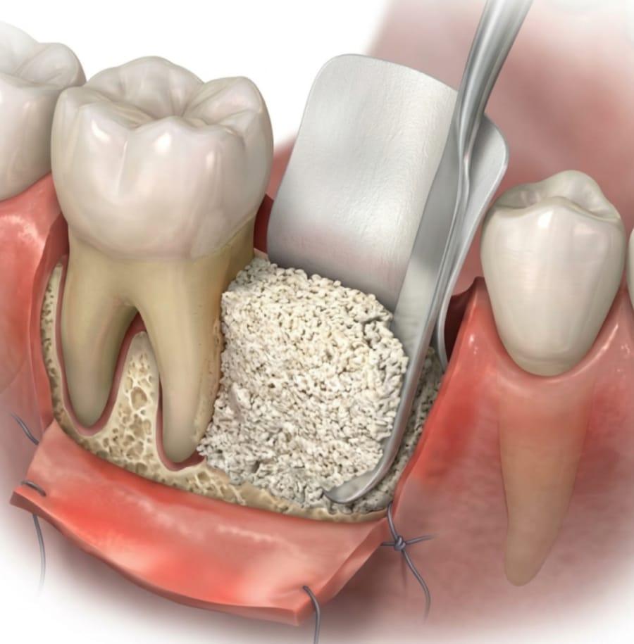
GBR rests on a simple principle: graft material is placed against the bone, then shielded from the gum by a membrane. The membrane gives the slower-growing bone cells time to fill the space before gum cells do. It can be resorbable (collagen) or not and, for larger reconstructions, reinforced with titanium or replaced by a titanium mesh that maintains the volume.
It is the most widely used technique for horizontal and moderate defects. For vertical gain, which is harder to achieve, a meta-analysis reports a mean gain of 4.2 mm with GBR versus 3.5 mm with bone blocks, and complications in 12% of cases with GBR versus 24% with blocks [3].
Bone blocks
A block is a piece of dense bone, most often taken from the patient’s own jaw (angle of the mandible, chin) or of animal origin, fixed to the ridge with screws. It is used mainly for defects in width. It often requires a second surgical site, with its own risks (sometimes temporary changes in sensation near the donor site).
Sinus lift
A sinus lift raises the thin membrane lining the sinus and places graft material beneath it, to create the bone height needed in the upper jaw behind the premolars. Two approaches exist, chosen according to the bone height remaining below the sinus.
- Lateral approach: through a small window in the sinus wall, when the remaining height is small. A systematic review reports 90.1% implant survival at three years; the most common complication is membrane perforation (19.5% on average), while graft infection is rare (2.9%) [4].
- Crestal (transalveolar) approach: through the implant site itself, when more height remains. Survival is 92.8% at three years, comparable to implants placed without grafting [5].
A membrane perforation noticed during surgery is most often repaired on the spot; it sometimes means postponing the graft.
Which materials?
| Material | Origin | Good to know |
|---|---|---|
| Autogenous | Patient’s own bone (chips or block) | Contains living cells; requires harvesting |
| Xenograft | Processed bovine bone with the organic part removed | Resorbs slowly and maintains volume; often mixed with autogenous bone |
| Allograft | Processed human bank bone | Avoids harvesting |
| Synthetic | Calcium phosphates | No biological origin |
For sinus lifts, a Cochrane review found no difference in failure between autogenous bone and bone substitutes, owing to insufficient evidence [6]. In practice, the choice depends on the volume to rebuild, the need to hold the shape over time, and your preferences.
What does a graft involve?
Whatever the technique, the graft is carried out under local anaesthetic, in the practice’s operating room in Levallois-Perret. The gum is lifted to expose the bone, the material is placed and protected (membrane, mesh or screws depending on the technique), and the gum is then closed without tension over the added volume. The grafted bone must then form before the implants are placed; a new cone beam CT, limited to the grafted area, is often used to check the volume obtained. The practical arrangements, the course of treatment and aftercare instructions are explained at the practice, according to your situation.
Risks, limitations and alternatives
Possible complications include exposure of the membrane or mesh through the gum, infection, partial loss of the graft, pain or swelling and, in the upper jaw, perforation of the sinus membrane. In the lower jaw, the nearby nerve makes 3D planning essential. A graft does not always yield all the volume hoped for: part of the material resorbs during healing, which is anticipated at the planning stage, and additional grafting is sometimes needed when the implant is placed.
Some situations reduce the chances of success or mean postponing surgery: smoking, poorly controlled diabetes, unstable periodontitis, thin or scarred gum that is hard to close, certain bone medications. The larger the volume to rebuild, especially in height, the higher the complication rate, as the figures quoted above show [3].
Grafting is not always the only option. In the lower jaw, a meta-analysis of randomised trials found comparable five-year results between short implants and vertical augmentation followed by standard implants, with fewer complications for short implants [7]. In the upper jaw, a Cochrane review could not separate sinus lift from short implants, but noted more complications with sinus lift [6]. A tooth-supported bridge or a removable denture also remain options, discussed according to your situation.
Frequently asked questions
Can the implant be placed at the same time as the graft?
Yes, when the remaining bone is enough to stabilise the implant and the defect is limited. For large defects, the graft comes first and the implant is placed once the new bone has formed.
Is animal bone used?
Often, yes: processed bovine bone is a widely studied material, sometimes mixed with your own bone. If you prefer to avoid it for personal or religious reasons, other materials can be discussed.
Can a graft fail?
Yes. Membrane exposure, infection or partial loss of the graft can occur. They sometimes require additional grafting. Smoking increases these risks.
How much does a bone graft cost?
It depends on the technique, the volume and the materials. The ranges charged at the practice are given on the Fees page, and a detailed estimate is provided after the assessment.
References
- Tan WL, Wong TL, Wong MC, Lang NP. A systematic review of post-extractional alveolar hard and soft tissue dimensional changes in humans. Clin Oral Implants Res. 2012;23 Suppl 5:1-21. DOI
- Atieh MA, Alsabeeha NHM, Payne AGT, Ali S, Faggion CM Jr, Esposito M. Interventions for replacing missing teeth: alveolar ridge preservation techniques for dental implant site development. Cochrane Database Syst Rev. 2021;4:CD010176. DOI
- Urban IA, Montero E, Monje A, Sanz-Sánchez I. Effectiveness of vertical ridge augmentation interventions: A systematic review and meta-analysis. J Clin Periodontol. 2019;46 Suppl 21:319-339. DOI
- Pjetursson BE, Tan WC, Zwahlen M, Lang NP. A systematic review of the success of sinus floor elevation and survival of implants inserted in combination with sinus floor elevation. Part I: lateral approach. J Clin Periodontol. 2008;35(8 Suppl):216-240. DOI
- Tan WC, Lang NP, Zwahlen M, Pjetursson BE. A systematic review of the success of sinus floor elevation and survival of implants inserted in combination with sinus floor elevation. Part II: transalveolar technique. J Clin Periodontol. 2008;35(8 Suppl):241-254. DOI
- Esposito M, Felice P, Worthington HV. Interventions for replacing missing teeth: augmentation procedures of the maxillary sinus. Cochrane Database Syst Rev. 2014;CD008397. DOI
- Terheyden H, Meijer GJ, Raghoebar GM. Vertical bone augmentation and regular implants versus short implants in the vertically deficient posterior mandible: a systematic review and meta-analysis of randomized studies. Int J Oral Maxillofac Surg. 2021;50(9):1249-1258. DOI
Patient pathway