Why plan in 3D before a bone graft and an implant?
A conventional X-ray shows bone from the front, like a shadow. It says nothing about its width. Yet width is often what decides whether an implant can stand on its own, whether grafting is needed, and how. Three-dimensional planning answers these questions before the gum is opened. Here is what it offers, and what it does not solve.
Key points
- Cone beam CT measures bone height and width, and locates the nerve in the lower jaw and the sinus.
- Planning starts from the future crown: the implant is placed where the tooth requires it, then the missing bone around it is measured.
- The plan is transferred to the mouth with a surgical guide and, for large reconstructions, with a titanium mesh designed on the same volume.
- Studies measure mean deviations of about 1 to 1.5 mm between plan and reality with a guide: a 2 mm safety margin is still needed.
- Limitations: X-ray dose, metal artefacts, accuracy that varies by machine, and a perfect plan does not ensure healing.
What cone beam CT shows, and a panoramic X-ray does not
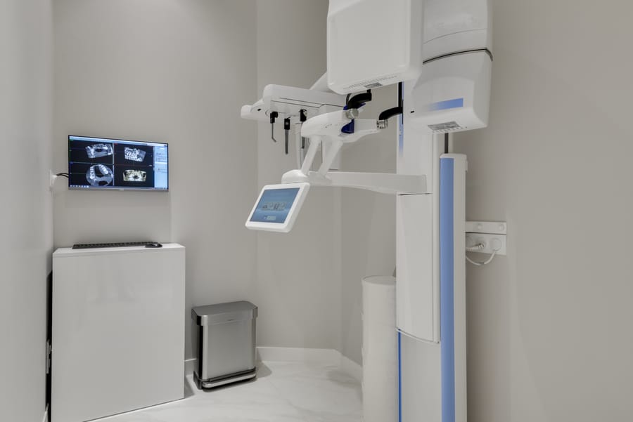
Cone beam CT (CBCT) reconstructs the jaws in three dimensions. The clinician can scroll through slices in every plane, including cross-sections perpendicular to the ridge that show the bone “in section”.
On these slices, the following are measured:
- the available bone height, from the top of the ridge to the nerve or sinus;
- the width of the ridge at several levels (a ridge can be wide at the base and knife-edged at the top);
- the angle of the ridge relative to the opposing teeth;
- the apparent density of the bone and any lesions (retained root, cyst, infection).
A review of recommendations considers cone beam CT justified for diagnosis, planning and intraoperative transfer in implant dentistry [1]. After an extraction, the ridge loses on average 3.8 mm of width in six months [2]: it is precisely this loss of width, invisible on a panoramic X-ray, that 3D imaging reveals.
Two areas to respect: the nerve and the sinus
In the lower jaw, the inferior alveolar nerve runs through the bone in a canal, below the roots of the premolars and molars. It supplies sensation to the lower lip and chin. Injury can cause loss of sensation or abnormal sensations. On the cone beam CT, the canal is identified and traced along its full length, together with its exit (the mental foramen) and any loops.
In the upper jaw, the sinus is an air-filled cavity lined with a thin membrane, just above the roots of the molars. Cone beam CT shows the bone height beneath the sinus, the thickness of the membrane, any internal walls (septa) and the condition of the sinus lining. These findings point towards a crestal or lateral sinus lift. During a lateral sinus lift, membrane perforation is the most common complication, occurring in 19.5% of cases on average in a systematic review [3]: knowing the anatomy before opening helps to anticipate it.
Planning from the tooth, not from the bone
For a long time, the implant was placed wherever bone remained, and the crown had to make do, sometimes at the cost of an oblique axis or a neck that was hard to clean. Prosthetically driven planning reverses the order of decisions. It proceeds in four steps:
- a digital impression of the teeth (taken with an intraoral scanner) is superimposed on the cone beam CT, so that the visible teeth and the invisible bone appear in the same space;
- the future crown is modelled in the place that chewing, appearance and cleaning require;
- the virtual implant is slipped beneath that crown, along the axis of chewing forces;
- only then is it checked whether bone surrounds this implant along its whole length. Where it is lacking, the volume to rebuild is defined around the implant as it should be, not the other way round.
For a periodontist, this logic matters in a particular way: an implant that is well aligned beneath its crown is easier to clean every day. Poor plaque control is one of the most firmly established risk factors for peri-implantitis [6]. Planning the position is therefore already preparing gum care around the implant.
Transferring the plan to the mouth: guide and mesh
A plan is only worth something if it is reproduced in theatre. Two tools act as a bridge between the computer and the mouth.
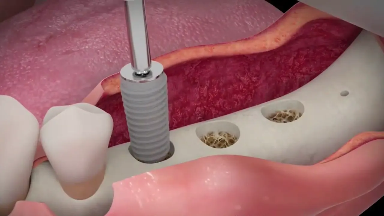
The surgical guide is a splint that directs the drills along the planned axis. Its accuracy has been measured: on average, the placed implant deviates from the plan by a little over a millimetre (1.2 mm at the entry point, 1.4 mm at the tip) and by 3.5° [4]. Guiding every step rather than only the first drill reduces these deviations further: compared with freehand placement, the angular deviation falls from 7.5° to 2.6° and the deviation at the tip from 2.2 mm to 0.9 mm [5]. The comparison table of the three techniques and the principle of placement are on the page Placing an implant and guided surgery.
For large reconstructions, the custom titanium mesh is designed on the same file: it gives physical form to the bone volume drawn around the implants. A 2025 meta-analysis reports, with these meshes, a mean horizontal bone gain of 6.4 mm and vertical gain of 5.1 mm [7]. Design and manufacturing are explained in the article on 3D printing, and the treatment itself on the Custom titanium mesh page.
What 3D does not solve: the limitations
- Dose. Cone beam CT delivers more radiation than a panoramic X-ray, and the dose varies widely between machines: from the equivalent of 2 to 200 panoramic images [1]. The field of view is therefore limited to what is needed, and the scan is only repeated if it will change the decision.
- Artefacts. Metal crowns, old implants or patient movement create streaks that hide the adjacent bone [1].
- Measurement accuracy. It can reach 0.2 mm on the highest-performing machines, but be several times worse depending on the machine and settings [1]. Figures read on screen are not measurements to the tenth of a millimetre.
- Transfer. Even with a guide, a deviation of about 1 to 1.5 mm remains on average [4]; the guide must be perfectly stable in the mouth.
- Biology. A perfect plan ensures neither gum healing nor bone formation. With custom meshes, exposure still occurs in about one case in five [7].
Finally, greater accuracy does not automatically translate into superior clinical outcomes: when the available systematic reviews are pooled, implant survival is the same after guided surgery and after freehand placement; the contribution of guidance would mainly be to reduce surgical risk in difficult cases [8]. 3D planning is a tool for decision-making and safety, not a promise.
In practice, when is 3D planning useful?
| Situation | Main contribution of 3D |
|---|---|
| Implant in the back of the lower jaw | Distance to the nerve, ridge width |
| Implant in the back of the upper jaw | Height below the sinus, choice of crestal or lateral sinus lift |
| Thin ridge after an old extraction | Width measurement, decision to graft |
| Large defect in height and width | Design of the volume to rebuild and of a custom mesh |
| Several implants or aesthetic zone | Positions and axes coordinated with the crowns |
In a simple situation with plenty of bone, planning is still useful but can be lighter. At Dr Hazout’s practice in Levallois-Perret, the level of planning is matched to the difficulty of the case: it is neither systematically maximal, nor reduced when the nerve or sinus are close.
Frequently asked questions
Is cone beam CT dangerous?
Its dose is higher than a panoramic X-ray but remains low when the field is limited to the area of interest. The scan is prescribed when it can change the treatment decision, which is the case before a graft or an implant.
Is a new cone beam CT needed after the graft?
Often yes, before the implants are placed, to check the bone volume obtained and adjust implant positions. The field is then limited to the grafted area.
Does the software decide instead of the surgeon?
No. It calculates and visualises; the clinician chooses the implant position, the grafting technique and the safety margins, taking into account the clinical examination and the gums.
Does a guide or a mesh make surgery risk-free?
No. They reduce the gap between plan and reality, but a deviation of about one millimetre remains, and healing also depends on the gums, smoking and oral hygiene.
References
- Jacobs R, Salmon B, Codari M, Hassan B, Bornstein MM. Cone beam computed tomography in implant dentistry: recommendations for clinical use. BMC Oral Health. 2018;18:88. DOI
- Tan WL, Wong TL, Wong MC, Lang NP. A systematic review of post-extractional alveolar hard and soft tissue dimensional changes in humans. Clin Oral Implants Res. 2012;23 Suppl 5:1-21. DOI
- Pjetursson BE, Tan WC, Zwahlen M, Lang NP. A systematic review of the success of sinus floor elevation and survival of implants inserted in combination with sinus floor elevation. Part I: lateral approach. J Clin Periodontol. 2008;35(8 Suppl):216-240. DOI
- Tahmaseb A, Wu V, Wismeijer D, Coucke W, Evans C. The accuracy of static computer-aided implant surgery: A systematic review and meta-analysis. Clin Oral Implants Res. 2018;29 Suppl 16:416-435. PubMed
- Werny JG, Frank K, Fan S, Sagheb K, Al-Nawas B, Narh CT, Schiegnitz E. Freehand vs. computer-aided implant surgery: a systematic review and meta-analysis—part 1: accuracy of planned and placed implant position. Int J Implant Dent. 2025;11:35. DOI
- Schwarz F, Derks J, Monje A, Wang HL. Peri-implantitis. J Periodontol. 2018;89 Suppl 1:S267-S290. DOI
- Ragucci GM, Fernández Augè A, Tresserra Parra A, Elnayef B, Hernández-Alfaro F. Comparison between CAD/CAM titanium mesh vs. conventional titanium mesh in bone regeneration: a systematic review and meta-analysis. Int J Implant Dent. 2025;11:55. DOI
- Tomar S, Chaudhary P, Ganguly A. Comparing the clinical outcomes of guided and freehand dental implant surgery: An umbrella review of systematic reviews and meta-analyses. J Prosthet Dent. 2026;135(5):e53-e59. DOI
Related treatments
Bone grafting before an implant: when, and which techniques
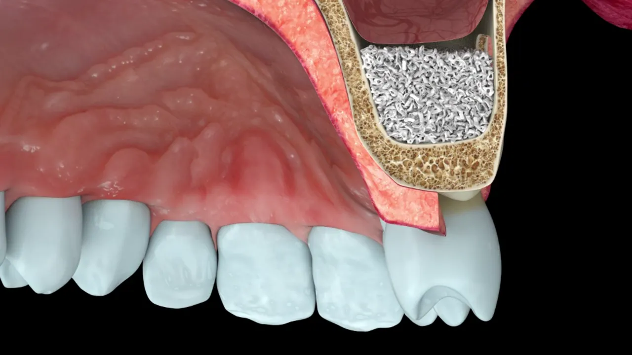
An implant must be surrounded by bone along its whole length. When bone is lacking, several techniques can rebuild it. They are not equally suited to every situation:…
Placing an implant: the steps, and what guided surgery adds
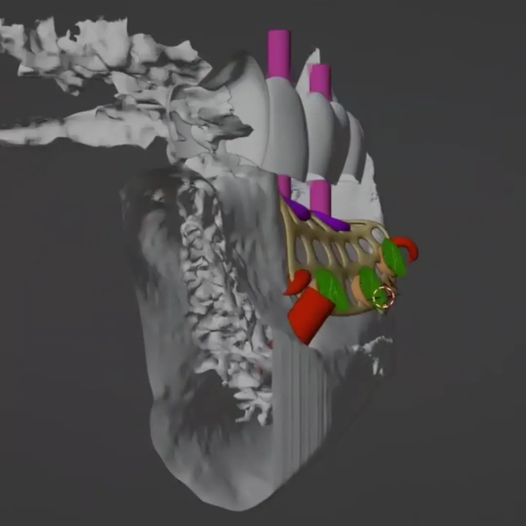
Implant placement is prepared well before the day of surgery. 3D imaging is used to decide where the implant should go; the surgical guide helps reproduce that decision…
Read next
How is a titanium mesh made by 3D printing?
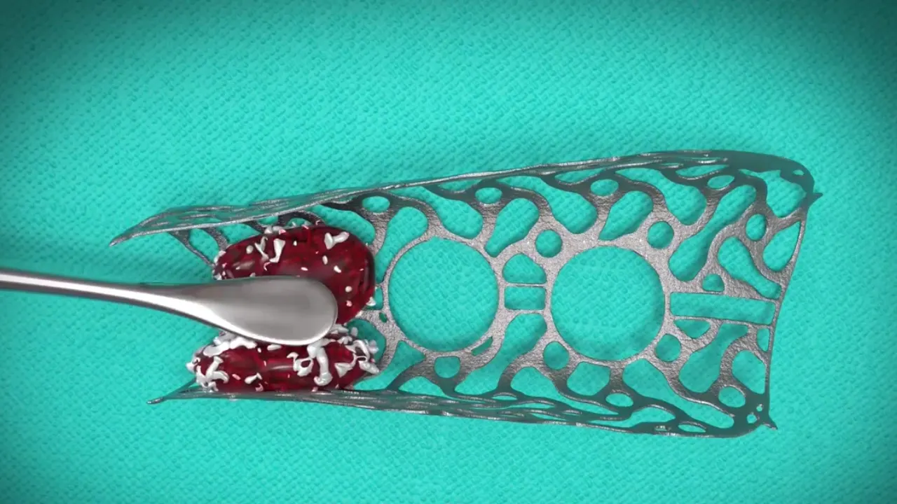
A custom titanium mesh is made as a single piece, for one patient and one bone defect. Between the scan image and the sterile part used in theatre lie a digital workflow…
Gum care around implants: preventing peri-implant mucositis and peri-implantitis
An implant cannot decay, but the gum and bone around it can become inflamed and break down, sometimes faster than around a tooth. The two diseases involved, peri-implant…
Patient pathway